
Fig. 7.
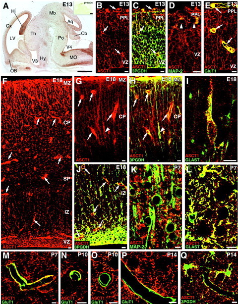
Cellular expression of ASCT1 in the developing cerebral cortex at E13 (A–E), E18 (F–J), P7 (K–M), P10 (N, O), and P14 (P, Q). A, Overview of ASCT1 immunostaining in the parasagittal brain section at E13. Intense ASCT1 staining is observed in the ventricular zone (VZ) and tubular profiles all over the brain wall. Theinset shows negative immunostaining with the use of preabsorbed antibody (preabs). B, C, Double immunofluorescence for ASCT1 (red) and 3PGDH (green). B, Single fluorescent image for ASCT1; C, merged view with 3PGDH.Arrows indicate tubular structures immunopositive for ASCT1 but not for 3PGDH. D, ASCT1 (red) and MAP-2 (green). MAP-2-positive neurons (arrowheads) in the preplate (PPL) are immunoreactive to ASCT1. E, ASCT1 (red) and GluT1 (green). Capillaries (arrows) are immunoreactive to both ASCT1 and GluT1.F, Single immunofluorescence for ASCT1 at E18. G, H, Double immunofluorescence for ASCT1 (red) and 3PGDH (green) in the superficial cortical region at E18. G, Single image for ASCT1; H, merged image with 3PGDH. Note intense fluorescence in radial fibers running in the cortical plate (CP) and marginal zone (MZ) as well as in capillaries (arrows). Also note a slender cell with radial fibers (arrowhead), which is immunoreactive to both ASCT1 and 3PGDH. I, ASCT1 (red) and GLAST (green). Such slender cells with radial fibers are immunoreactive to GLAST, indicating migrating radial glia cells or astrocytes. J, ASCT1 (red) and 3PGDH (green) in the deep region of the cortex. Note intense double fluorescence in neuroepithelial cells of the ventricular zone (VZ) and in dispersed cells of the intermediate zone (IZ).K, Nonoverlapping pattern for ASCT1 (red) and MAP-2 (green) at P7. L, Extensive costaining for ASCT1 (red) and GLAST (green) at P7. M–P, Double immunofluorescence for ASCT1 (red) and GluT1 (green) showing the loss of capillary expression of ASCT1 during the second postnatal week. At P7, most capillaries are immunoreactive to both ASCT1 and GluT1 (M). At P10, two types of capillaries are observed, one retaining ASCT1 expression (N) and the other lacking ASCT1 (O). At P14, many capillaries are negative to ASCT1 (P).Q, ASCT1 (red) and 3PGDH (green) at P14. ASCT1 and 3PGDH are well overlapped in astrocytes and also in their processes surrounding capillaries. Hy, Hypothalamus; SP, subplate; Th, thalamus. See other abbreviations in the legend to Figure 1. Scale bars: A, 0.5 mm;B–Q, 20 μm.