
Fig. 8.
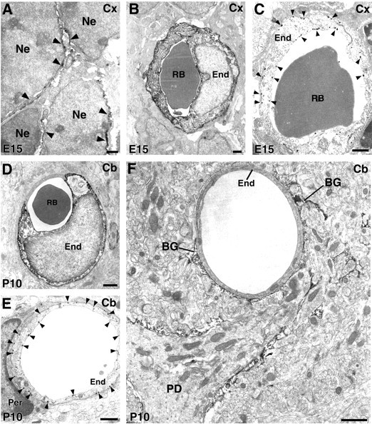
Immunoelectron microscopy for ASCT1 in the developing cerebral cortex at E15 (A–C) and cerebellar cortex at P10 (D–F).A, ASCT1 labeling is detected in the surface (arrowheads) of neuroepithelial cells (Ne). B, Immunoperoxidase labeling for endothelial cells (End) of cortical capillaries at E15.C, Immunogold labels the luminal and abluminal cell membranes of endothelial cells (arrowheads).D, Immunoperoxidase labeling of endothelial cells of cerebellar capillaries at P10. E, Immunogold labels the luminal and abluminal cell membranes of endothelial cells (arrowheads). F, Some capillaries at P10 lose ASCT1 expression and are surrounded by Bergmann glia processes (BG) with intense ASCT1 labeling. Per, Pericyte; RB, red blood cell. Scale bars, 1 μm.