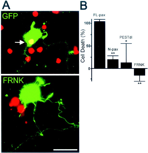
Fig. 6.
PTP-PEST and FAK activity are involved in Aβ-induced neuronal death. A, Aβ treatment induces cell death in neurons expressing GFP (top panel, greenfluorescence). Nonviable cells show process retraction and disintegration and positive nuclear staining for propidium iodide (arrow). Neurons expressing FRNK (bottom panel, green fluorescence) became dystrophic but remained viable. Note the dystrophic appearance of neuritic processes and the absence of propidium nuclear staining in the neuron expressing FRNK.B, Quantification of cell death in transfected neurons treated with Aβ. A significant reduction in Aβ-induced neuronal death is observed in neurons expressing N-pax, which lacks paxillin LIM domains (20.4 ± 8%), andPESTdl, which lacks the paxillin-binding domain of PTP-PEST (13.3 ± 41.7%). FRNK completely prevents neuronal death (−15.3 ± 13%). Cell death is expressed as a percentage of Aβ-induced propidium-positive neurons transfected with GFP (100%). Values are mean ± SEM; n = 3 independent experiments; >200 neurons were scored per condition in each individual experiment; *p < 0.05; **p < 0.01 relative to control (GFP) by ANOVA followed by the Student–Newman–Keuls post hoc test. Cortical neurons were transfected at day 5, treated with fibrillar Aβ for 2 d, and processed for analysis. Before fixation, the nuclei of dead cells were labeled with propidium iodide.

