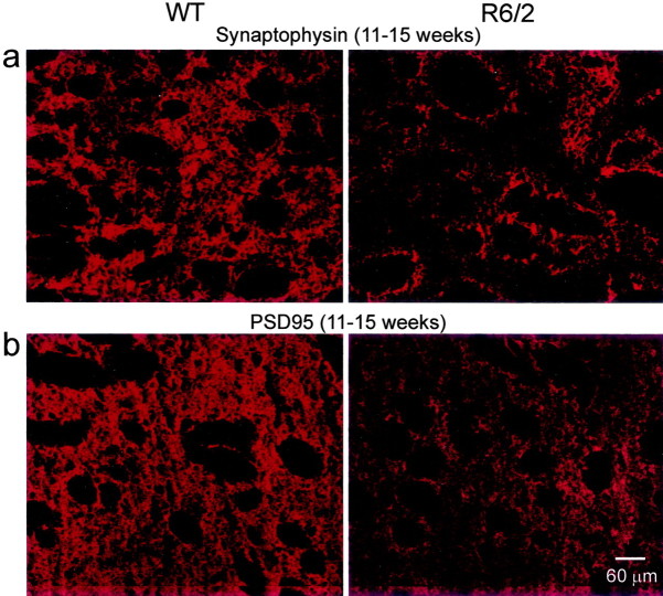
Fig. 7.
a, Synaptophysin staining was visible throughout the presynaptic cytoplasmic compartments of afferent inputs and locally derived endings in the WT striatum at 11–15 weeks (left) but was attenuated considerably in the R6/2, corresponding to the loss of the presynaptic compartment at 11–15 weeks (right). Fiber bundles that penetrate the striatum appeared as black, nonstained myelinated structures in botha and b. b, Expression of PSD95 was detected within the striatal neuropil of the WT at 11–15 weeks (left) but was reduced substantially at 11–15 weeks in the R6/2 (right). Scale bar refers to allpanels.

