
Fig. 3.
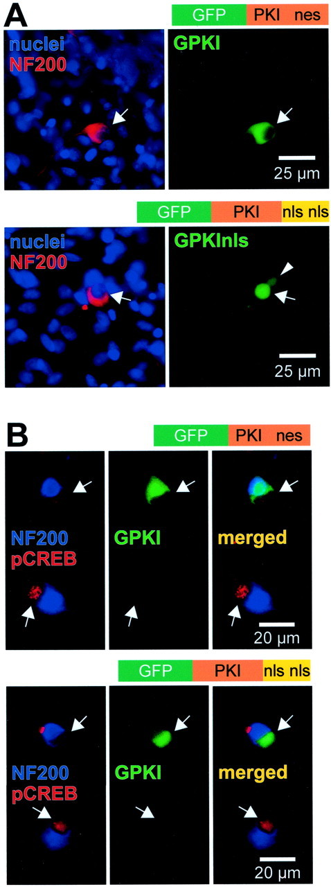
Expression and function of subcellularly targeted GFP-tagged PKI constructs in SGNs. SGNs were transfected with GPKI, which contains an intrinsic nuclear export signal, or with GPKInls, in which the nuclear export signal was deleted and replaced with two nuclear localization signals, as indicated. A, After allowing 24 hr for expression, the cultures were fixed, and nuclei and neuronal cytoplasm were labeled with Hoechst 33342 and anti-NF200, respec- tively, as in Figure 1. The diagrams above each pair of images show the general structure of the construct transfected. Theleft panel of each pair shows a composite of the nuclei and NF200 images. The corresponding GPKI or GPKInls images are shown atright. The arrows identify the location of neuronal nuclei and indicate identical positions in bothpanels of each pair. The arrowheadindicates the nucleus of a fortuitously transfected glial cell. Green fluorescence is evident primarily in the cytoplasm of the GPKI-transfected neuron and primarily in the nucleus of the GPKInls-transfected neuron. B, Cultures transfected with GPKI (top) or GPKInls (bottom), as inA, were treated for 30 min with 1 mmcpt-cAMP before fixation. Two neighboring SGNs are shown in each set ofpanels, one (top) transfected and one (bottom) untransfected. In each set,left, superimposed blue (NF200) and red (phospho-CREB) images identifying SGNs and showing phospho-CREB immunoreactivity.Center, Same field using a green filter to detect GFP and shows the upper neuron of each pair expressing the indicated GFP-tagged PKI construct. Right, Mergedleft and center images. For either construct, the transfected neuron exhibits greatly reduced phospho-CREB immunoreactivity relative to the untransfected.