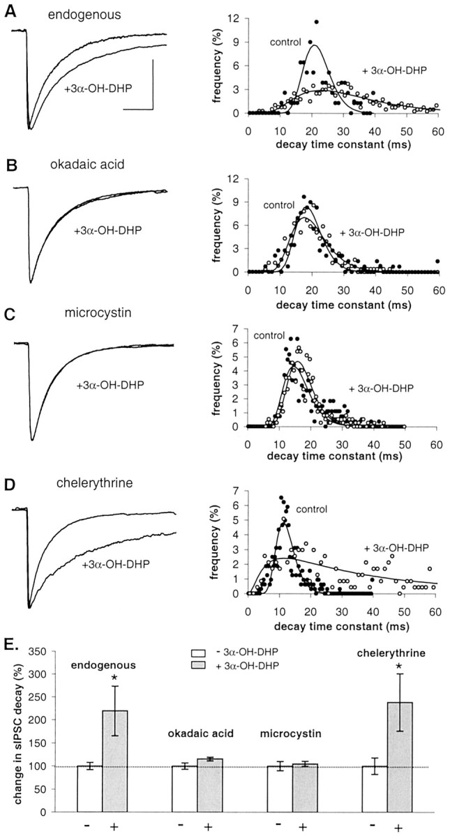
Fig. 4.

Effect of 3α-OH-DHP is dependent on Ser/Thr phosphatase activity in juvenile male rats (postnatal days 21–27). B, C, Average sIPSCs in the absence and presence of 3α-OH-DHP and decay time constant histogram showing that inhibition of phosphatase activity by 50 nm okadaic acid (A) or 100 nm microcystin (B) prevents the effect of 3α-OH-DHP. D, Average sIPSCs and histogram of sIPSC decay time constants obtained in the presence of 1 μm chelerythrine showing that PKC inhibition does not prevent the effect of 3α-OH-DHP. E, Summary graph illustrating the relative effects of 3α-OH-DHP on sIPSC decay time constants in the four experimental conditions. Decay plotted as percentage of control values. White bars, Controls;gray bars, 3α-OH-DHP. Relative neurosteroid effects are as follows: endogenous condition, 220 ± 54% of control,p < 0.05, n = 7; 50 nm okadaic acid, 115 ± 4% of control,p > 0.05, n = 7; 100 nm microcystin, 105 ± 6% of control,p > 0.05, n = 5; 1 μm chelerythrine, 239 ± 63% of control,p < 0.05, n = 5. Okadaic acid (104 ± 3%; n = 7), microcystin (97 ± 2%; n = 10), and chelerythrine (99 ± 8%;n = 5) by themselves had no effect on sIPSC decay (unpaired t test; p > 0.05). In none of these conditions was the sIPSC amplitude significantly affected (data not shown). All other traces of average sIPSCs were plotted normalized to the control average in A. Calibration (of the control trace), 20 msec, 100 pA.
