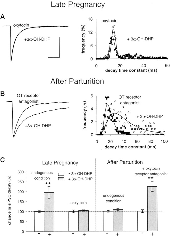
Fig. 7.
Oxytocin autoregulation renders GABAARs insensitive to neurosteroid. A,Left, Superimposed average sIPSCs before and after 3α-OH-DHP application obtained in SON neurons at P20 pretreated with OT. Right, Decay time constant histogram of this experiment. B, Average sIPSCs in the absence and presence of 3α-OH-DHP and decay time constant histogram showing large 3α-OH-DHP effect at PPD1 after block of oxytocin autoreceptors.C, Summary graph illustrating the dependence of the neurosteroid effect on oxytocin receptor activity. Decay plotted as percentage of controls. White bars, Control; gray bars, 3α-OH-DHP. Notice the differences with the endogenous condition at both stages. After OT pretreatment, 3α-OH-DHP did not affect sIPSC decay time constants (104 ± 5% compared with during OT pretreatment; p > 0.05; n = 6). OT by itself had a small but nonsignificant effect on sIPSC decay time constants (114 ± 7% of control; p > 0.05; n = 8). As expected, OT decreased sIPSC amplitudes (68 ± 13% of control; p < 0.05;n = 8). At PPD1, 3α-OH-DHP increased sIPSC decay time constants after OT antagonist (224 ± 26% compared with during OT antagonist pretreatment; p < 0.05;n = 5). OT antagonist by itself had no effect on sIPSC decay (109 ± 5% of control; p > 0.05;n = 5). OT antagonist had no effect on sIPSC amplitude (96 ± 19%; n = 5). All other traces of average sIPSCs were plotted normalized to the control average in A. Calibration (of the control trace), 20 msec, 100 pA.

