
Fig. 2.
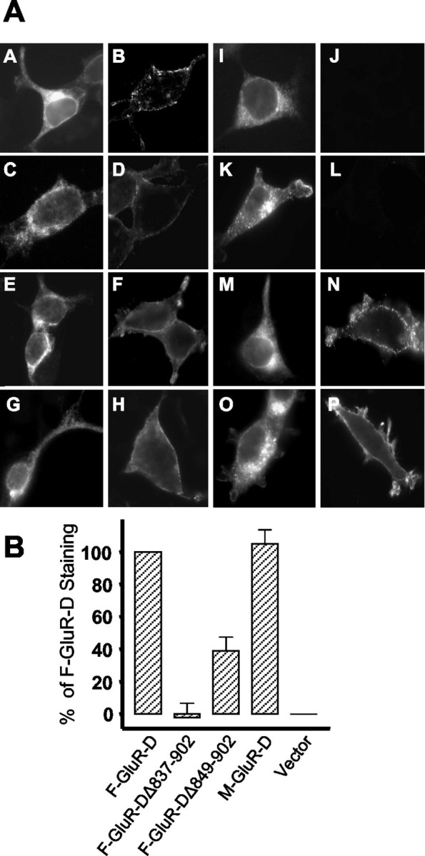
Surface expression in transfected HEK293 cells.A, Immunostaining of GluR-D constructs after transfection of HEK293 cells; A, B, nontagged GluR-D; C, D, Flag-GluR-D;E, F, Myc-GluR-D; G,H, GluR-D-His; I, J, GluR-DΔ837–902; K, L, GluR-DΔ841–897; M, N, GluR-DΔ849–902; O, P, GluR-DΔ897–902. A and B were probed with Fab 7 IgG; E and F were probed with anti-Myc IgG; C, D, G–Pwere probed with anti-Flag IgG. Permeable staining is shown inpanels A, C, E,G, I, K, M, and O. Nonpermeabilized expression is shown inpanels B, D, F,H, J, L, N, and P. B, Quantitation of the degree of cell-surface expression of the GluR-D deletion mutants by ELISA. They-axis indicates the level of surface expression as a percentage of the full-length, Flag-tagged GluR-D construct. The values were calculated from the A405 readings (see Materials and Methods). Four separate transfections were done of all the constructs in parallel. The values were corrected for nonspecific labeling.