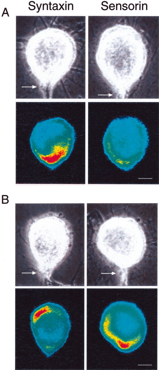
Fig. 1.

Interaction with L7 leads to opposite changes in the distribution of syntaxin mRNA versus sensorin mRNA.A, Syntaxin mRNA accumulates at the axon hillock on day 1, whereas sensorin mRNA is distributed uniformly. The top micrographs are Nomarski contrast images of each SN cell body with its main axon (arrow) emerging at the 6 o'clock position. The bottom micrographs are pseudocolor representation of fluorescent signals (red is high intensity; blue is low intensity) for syntaxin or sensorin mRNA in SNs cultured with L7 for 1 d. Neurites around SN cell bodies belong to both SN and L7. Scale bar, 25 μm.B, Syntaxin mRNA accumulates away from the axon hillock, but sensorin mRNA accumulates at the axon hillock on day 5. Thetop micrographs are Nomarski contrast views of each SN cell body and axon; the bottom micrographs are pseudocolor representations of fluorescent signals for syntaxin and sensorin mRNAs in the SNs cultured with L7 for 5 d. Scale bar, 25 μm.
