
Fig. 6.
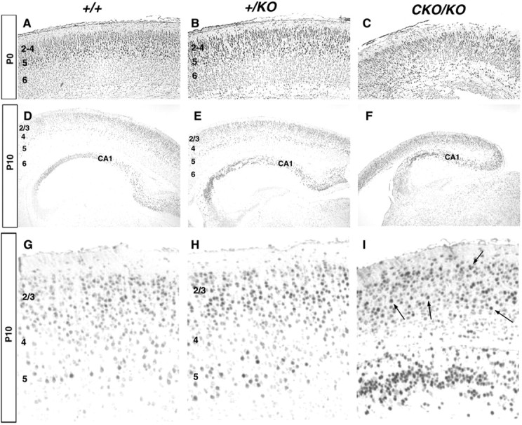
Tst-1 immunohistochemistry on caudal coronal sections. A–C, P0 brains. StrongTst-1 expression was present in layers 2–4 and 5 in both wild-type (A) and+/KO (B) brains with a slightly broader layer 5 in +/KO. Expression was diffusely detected throughout the cortex in CKO/KO(C). D–F, P10 brains, with higher magnification in G,H, and I. Expression was confined mainly to cortical layers 2–3 and 5 in both wild type (D, G) and +/KO (E, H). In theCKO/KO brains (F, I), labeled neurons were scattered throughout the cortex (arrows), with no obvious lamination pattern. Note the strongTst-1 expression in CA1 of the hippocampus in all genotypes.