
Fig. 4.
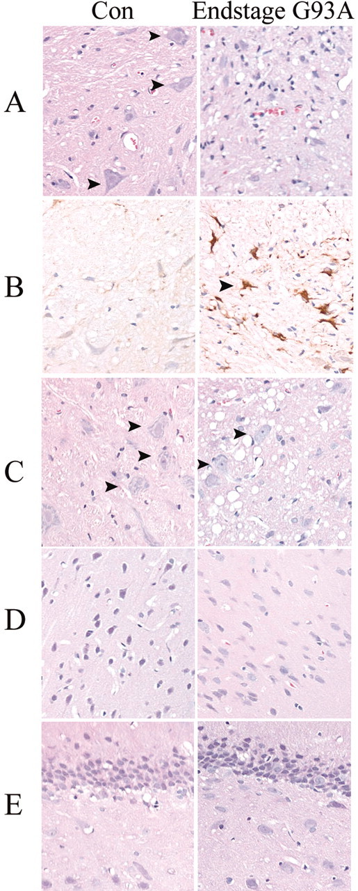
Histochemical analysis of pathology in end-stage G93A transgenic rat spinal cord and brain regions. Cervical spinal cord (A) or brain areas including brainstem (C), neocortex (D), and hippocampus (E) were stained with hematoxylin and eosin to show extent of neuronal loss, degenerative structures (vacuoles), as well as gliosis in end-stage G93A rats compared with age-matched control rats (Con). Arrows inA and C denote the presence of large motor neuron cell bodies, rarely found in end-stage diseased spinal cord. Representative spinal cord sections from control and end-stage G93A rats are also shown stained with anti-GFAP antibody (B). Arrows in Bhighlight a hypertrophic astrocyte, frequently found in end-stage diseased spinal cord.