
Fig. 9.
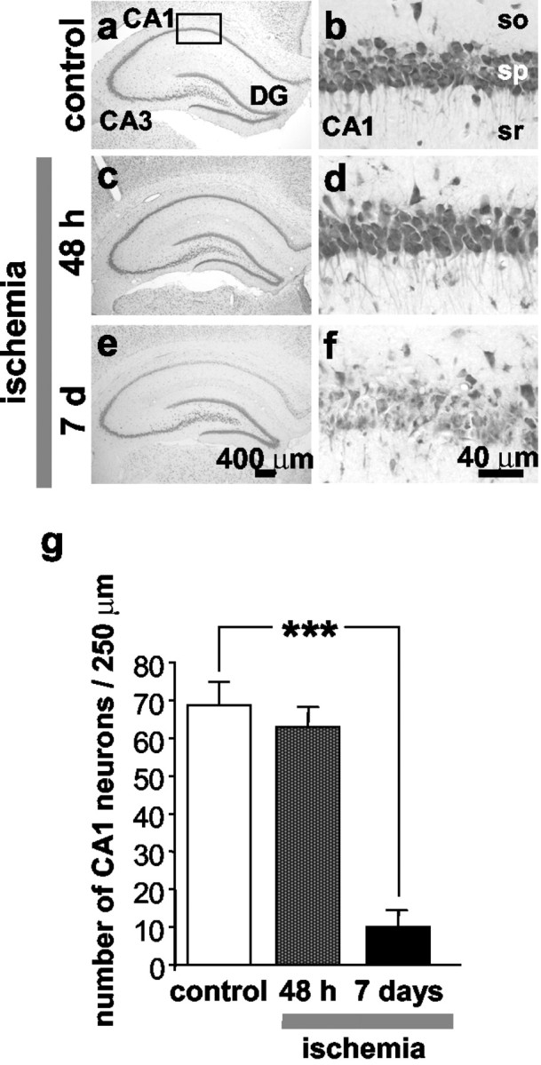
Global ischemia induces selective, delayed neuronal death in hippocampal CA1. a–f, Toluidine blue staining of coronal brain sections at the level of the dorsal hippocampus from control (a, b) (n = 5) and experimental male rats subjected to global ischemia at 48 hr (c, d) (n = 9) and 7 d (e,f) (n = 5) after ischemia. Control animals were killed at 7 d after sham operation. At 48 hr after global ischemia, there was no histologically detectable neuronal death in any hippocampal subfield. At 7 d after ischemia, the pyramidal cell layer of CA1 exhibited dramatic loss of neurons, whereas CA3 and dentate gyrus showed no damage. Scale bars: a,c, e, 400 μm; b,d, f, 40 μm. g, Quantitation of cell counts from brain sections illustrated ina–f. To assess hippocampal injury, the number of surviving neurons per 250 μm length in the pyramidal cell layer of the medial CA1 was counted under a light microscope at 40× magnification in sections. Neuronal counts from a minimum of four microscopic sections per animal were analyzed; comparisons among group means were made using the Student's t test (***p < 0.001).