
Fig. 5.
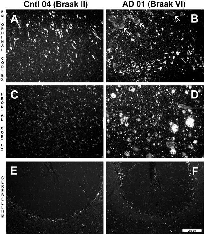
Paraffin sections (8 μm) from the entorhinal cortex (A, B), frontal cortex (C, D), and cerebellum (E,F) of a Braak stage II control brain (A, C, E, Cntl 04) and a Braak stage VI AD brain (B,D, F, AD 01) stained with X-34. X-34 is a highly sensitive, fluorescent Congo red derivative that intensely stains plaques, NFTs, and amyloid deposits in general (Styren et al., 2000). Marked atrophy of AD tissue was notable in all regions. In the entorhinal cortex of the control subject (A), frequent numbers of NFTs were seen, whereas there were no amyloid deposits. In the AD case (B), there also were frequent NFTs along with diffuse amyloid deposits (arrows). X-34-positive NFTs and amyloid plaques were absent from the frontal cortex of the control case (C), whereas they were abundant in AD (D). There was no detectable X-34 staining of plaques or NFT in control (E) and AD (F) cerebellum. Control frontal cortex (C) and cerebellum from both control and AD (E, F) show small stellate cells that stain with X-34. Scale bar, 200 μm.