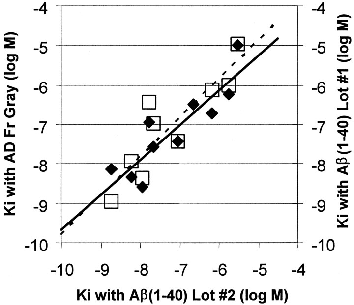
Fig. 6.
Comparison of the Kivalues of 10 BTA-1 derivatives for inhibition of [3H]BTA-1 binding to Aβ(1–40) fibrils and homogenates of AD frontal gray (filled diamonds,solid line). Also shown is a comparison of the same data for Aβ(1–40) fibrils compared with Kivalues previously determined in a separate, older lot of Aβ(1–40) fibrils (open squares, dashed line).

