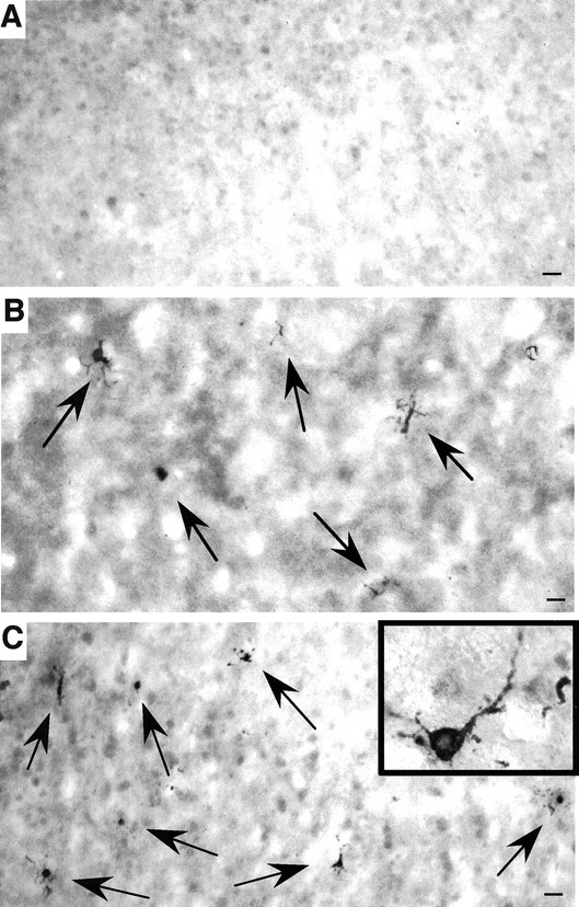
Fig. 3.

Caspase-3-activated neurons in N171-82Q mouse brain. The cortex of a wild-type control mouse at 14 months of age (A) and the striatum (B) and cortex (C) of N171-82Q mice at 4.5 months of age were stained with an antibody against the activated form of caspase-3. Positively stained neurons (arrows) are present in the N171-82Q mouse brain regions. Inset, High-magnification micrograph of a neuron containing activated caspase-3. Scale bars, 32 μm.
