
Fig. 7.
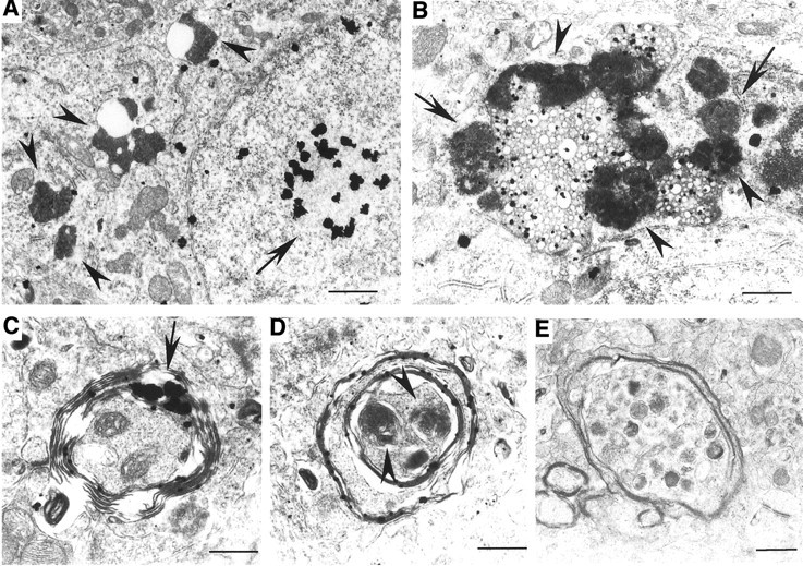
Degenerated neurons and axons in Hdh150CAG knock-in mice. A, B, Cytoplasmic organellar degeneration in HdhCAG150 mouse striatum. A, Several dark cytoplasmic organelles surround vacuoles (arrowheads). The nucleus that contains an intranuclear inclusion (arrow) appears intact, without nuclear fragmentation or condensation.B, A high-magnification micrograph shows clusters of electron-dense bodies and dark lysosomal structures that surround many cytoplasmic vacuoles. Some of these dense bodies show a mitochondrial origin with internal cristas (arrows). Others may represent secondary lysosomal bodies (arrowheads).C–E, Various types of degenerating axons are present in the striatum of HdhCAG150 mice. Axons are surrounded by disrupted myelin in which huntingtin aggregates (arrow) are trapped (C). Degenerated axons contain dark and swollen organelles (D, E), probably derived from mitochondria (arrowheads in D). Scale bars: A, 1 μm; B, 0.63 μm;C, 0.42 μm; D, 0.41 μm;E, 0.71 μm.