
Fig. 3.
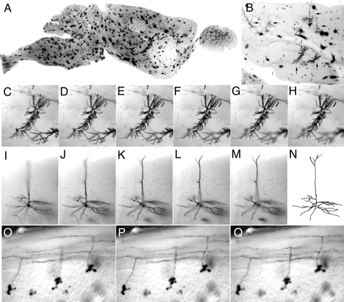
Morphology of adult CNS neurons. Brain from an adult R26CreER/+;ZAP/+ mouse that received a single intraperitoneal injection of 0.8 mg of 4HT at P21. A, Parasagittal section (300 μm) through the brain. Most of the intensely labeled cells are glia. B, Section (300 μm) through cortex (top) and hippocampus (center) reveals labeled pyramidal neurons (horizontal arrows) and CA1 neurons (vertical arrows). C–H, Successive focal planes through one of the CA1 neurons shown inB. I–M, Successive focal planes through a pyramidal neuron and its reconstructed image (N). O–Q, Three cerebellar granule cells. The cell bodies and compact dendritic trees are seen in the bottom half of each panel; the characteristic T-shaped branching of the axon that gives rise to parallel fibers is seen in the top half of each panel. In C–Q, successive focal planes are at intervals of 5 μm.