
Fig. 4.
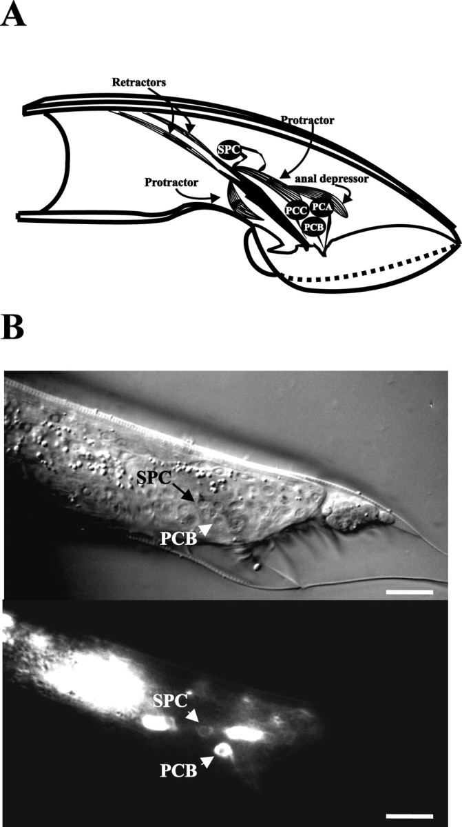
Cells involved inunc-103(sy557)-induced spicule protraction. A depicts the right lateral view of the male tail, highlighting cells that are associated with the spicules. The drawing was adapted from Sulston et al. (1980). The spicule is shown in black. The dorsal and ventral protractor muscles, represented as striated cells, are attached to the body wall and the base of the spicule. The anal depressor muscle is attached to the body wall and contacts the dorsal protractor. The right SPC motor neuron contacts the protractors and the base of the spicule. The right PCA, PCB, and PCC neurons send their sensory processes to the right postcloacal sensillum. B depicts Nomarski (top) and fluorescence (bottom) images of the lateral tail region of a male that expresses theunc-103:: GFP reporter construct. Scale bars, 20 μm. The male in the image is in L4 lethargus. The PCB postcloacal sensory neuron and the SPC spicule motor neuron, in addition to some unidentified neurons, express the reporter gene.