Fig. 5.
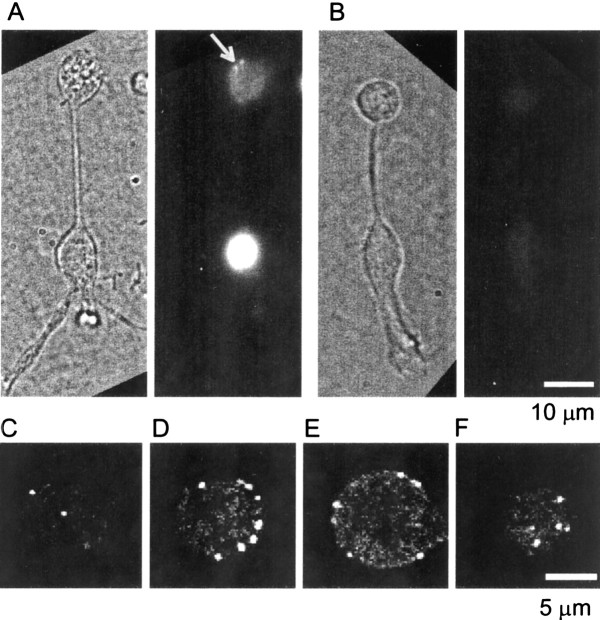
Immunofluorescent localization of synaptic ribbons in bipolar cells. A chemically fixed single-dissociated bipolar cell is shown, immunostained with an antibody that recognizes CtBP1, CtBP2, and the B-domain of the ribbon protein ribeye. A, Active serum preincubated with uncoated nickel beads. Left, Bright field; right, fluorescence; arrowpoints to two ribbons. B, Image as inA but with serum immunodepleted by preincubation with nickel beads coated with recombinant His-tagged CtBP2.C–F, Confocal sections taken from the terminal of another bipolar cell, stained as described above except that the preincubation was omitted. Panels show the bottom-most section (C), two additional sections 1 μm (D) and 4 μm (E) higher, and the top-most section (F). Note that, except in the bottom and top sections, spots are found on the perimeter of the terminal, indicating that most or all staining is on the surface.

