Fig. 1.
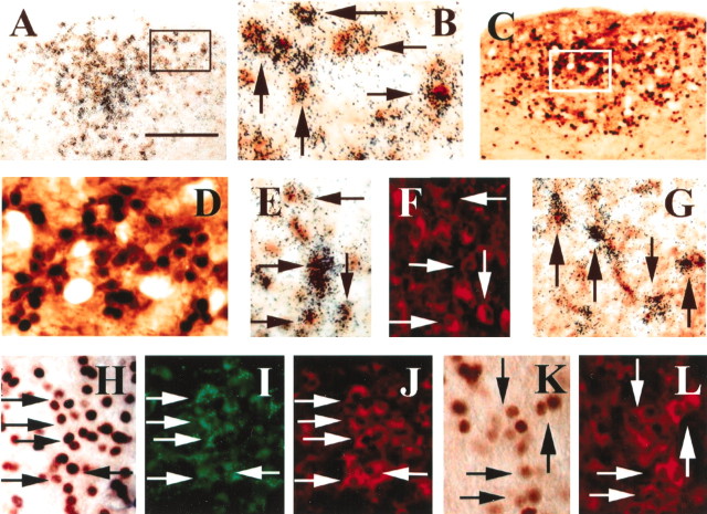
Intravenous EXN-4 activated neurons in the AP.A, B, In situhybridization coupled with immunohistochemistry demonstrates TH-immunoreactive neurons expressing GLP-1R in the AP. The neurons containing clusters of silver grains were hybridized with a GLP-1R35S-labeled riboprobe. The TH-immunoreactive neurons contain brown cytoplasmic reaction product. B is a higher magnification of A (arrowsindicate double-labeled neurons). C, D, Dual-label immunohistochemistry demonstrates that TH-immunoreactive neurons (brown cytoplasm) contain Fos-IR (black nuclei) 2 hr after intravenous EXN-4 in the AP.D is a higher magnification of C.E, F, In situhybridization coupled with dual-label immunohistochemistry demonstrates that many GLP-1R-expressing TH-immunoreactive neurons (red neurons in F) are activated by intravenous EXN-4. The neurons containing clusters of silver grains were hybridized with a GLP-1R 35S-labeled riboprobe, and the Fos-immunoreactive neurons contain brown cytoplasmic reaction product in E (arrows indicate triple-labeled neurons). G, In situhybridization coupled with immunohistochemistry demonstrates that many retrogradely labeled neurons (brown cytoplasm) after FG injection into the PBel also express GLP-1R mRNA (clusters of silver grains) (arrows indicate double-labeled neurons).H–J, Triple-label immunohistochemistry reveals that many retrogradely labeled neurons (green neurons inI) in the AP contain TH-IR (redcytoplasm in J) and also contain Fos-IR (brown nuclei in H) after intravenous EXN-4 (arrows indicate triple-labeled neurons). K, L, Dual immunohistochemistry demonstrates that TH-immunoreactive neurons (redcytoplasm in L) also contain intravenous albumin-conjugated GLP-1-induced Fos-IR (brown nuclei inK) (arrows indicate double-labeled neurons). Scale bar: A, C, 200 μm;B, D, E–L, 50 μm.

