
Fig. 4.
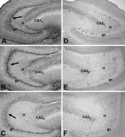
Seizure-induced reduction of GAT-1 and CCK immunoreactivity in the dentate gyrus of kindled rats experiencing spontaneous seizures. Immunoreactivity was intensified in these sections by silver treatment, as described in Materials and Methods, and all sections were batch processed. A, In normal rats, GAT-1 immunoreactivity is observed throughout the neuropil, but is particularly prominent in the granule cell layer (gc) bordering the subgranular region of the hilus (H;arrows), which is the site of axon initial segments arising from granule cells and projecting into the hilus toward CA3c. B, In a kindled rat that experienced 76 class V seizures, which is a stage just before the onset of spontaneous seizures, GAT-1 immunoreactivity appeared normal in the dentate gyrus. C, In a kindled rat that experienced 209 evoked class V seizures, immunoreactivity was reduced in comparison with normal or age-matched controls. The loss of GAT-1 immunoreactivity was particularly prominent along the border of the granule cell layer and subgranular region of the hilus, which is the site of axon initial segments and spike initiation. D, In a nearby section of the dentate gyrus from the normal rat in A, GABAergic interneurons labeled by CCK were located along the subgranular region near the border of the granule cell layer and the hilus and were occasionally also observed deeper in the hilus. Fine punctate granules labeled by CCK were also typically scattered throughout the hilus and granule cell layer in normal rats. E, In the same kindled rats that experienced 76 class V seizures, the distribution and number of CCK-labeled interneurons and punctate granular staining appeared normal. F, In the same kindled rat shown inC that experienced 209 evoked class V seizures, there was a reduction of interneurons labeled by CCK and in the punctate staining observed in the hilus and granule cell layer. Scale bar, 200 μm.