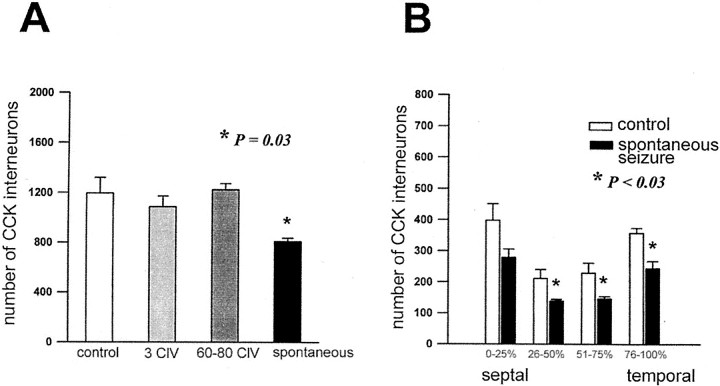
Fig. 5.
Reduction of CCK-immunoreactive GABAergic interneurons in the dentate gyrus of kindled rats experiencing spontaneous seizures. The number of CCK-immunoreactive interneurons in the dentate gyrus was assessed as a function of the number of evoked seizures and was compared with normal controls matched in age to kindled rats that experienced >100 class V seizures. Profile counts of CCK-immunoreactive interneurons were counted in the dentate gyrus by stereological methods in consecutive transverse sections from the septal to temporal pole of the extended hippocampus (see Materials and Methods for details). A, There was a significant reduction of CCK-immunoreactive interneurons in kindled rats with >100 class V (ClV) seizures (filled bar) compared with age-matched normal controls (open bar) and kindled rats examined after 3 or 60–80 evoked class V seizures (gray bars). B, In kindled rats with >100 evoked class V seizures (filled bars), reduction of CCK-immunoreactive interneurons was observed along the entire septotemporal axis of the hippocampus. Percentages of 0–25, 25–50, 50–75, and 75–100 refer to relative location along the septotemporal axis. There were significant reductions of CCK-immunoreactive interneurons from the middle through the most temporal extent of the dentate gyrus and a trend toward reduction at the most septal location. Asterisks indicate significant differences.

