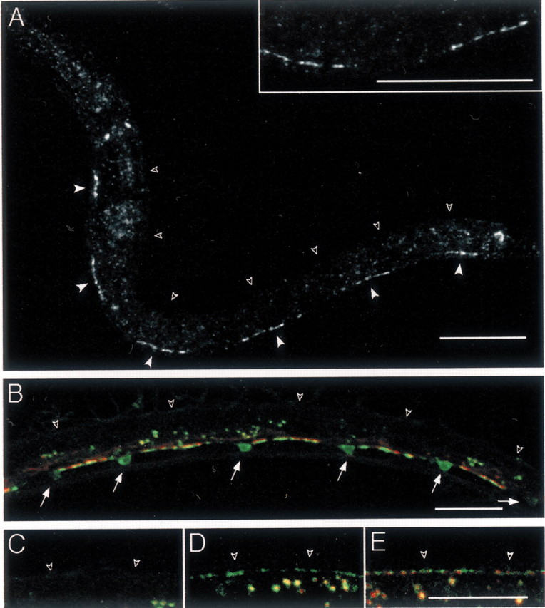
Fig. 2.

GABA receptor clusters are present only at sites of innervation in L1 larvae. A, Immunostaining of UNC-49 in wild-type L1 larvae. L1 larvae were immunolabeled 4 hr after hatching at 25°C. At this stage, UNC-49 was found ventrally as six segments (closed arrowheads) interrupted by gaps; however, no staining could be observed in the dorsal nerve cord (open arrowheads). Inset shows a higher magnification of the ventral nerve cord revealing that UNC-49 receptors form clusters. B, SNB-1–CFP (in green) expressed in GABAergic neurons stains synaptic vesicle clusters present along the ventral nerve cord and the soma of the six GABAergic DD motor neurons present in young L1 larvae (arrows). Clusters of UNC-49–YFP (red) are detected only along the ventral nerve cord. Open arrowheads indicate dorsal cord position. The fluorescence in the middle of the animal corresponds to autofluorescent gut granules. C–E, Dorsal cord labeling (open arrowheads) in representative larvae grown at 25°C after egg laying for 16 hr (L1 larval stage) (C), 24 hr (early L2 stage) (D), and 26 hr (L2 larva) (E). Presynaptic vesicles (labeled with SNB-1–CFP in green) are detected before UNC-49–YFP receptor clusters (red). Scale bars, 20 μm (for all images).
