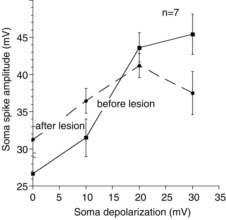
Fig. 9.
Afferent spikes in the soma before and after the lateral process lesions. When neurons were at their resting membrane potential, afferent spikes recorded after the lateral process had been lesioned were larger than spikes recorded before the lateral process was lesioned (two-tailed paired t test;p = 0.01) (a significant result even when a Bonferroni correction for the repeated measures is applied). When neurons were progressively depolarized, spike amplitude increased both with and without the lateral process (two-factor repeated-measures ANOVA; p < 0.001 for membrane potential;p = 0.0027 for membrane potential and lesion status). Note that, although spikes were initially smaller before the lesion, spike amplitude was increased at least as much as it was after the lesion (10, 20, and 30 mV spike amplitudes are not statistically different when a Bonferroni correction is applied). Overall, therefore, the effect of the lesion was not statistically significant (two-factor repeated-measures ANOVA; p = 0.87).

