
Fig. 6.
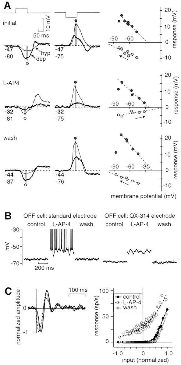
The ON pathway inhibits an OFF cell phasically to a light flash and tonically at mean luminance. A, The response of an OFF cell was measured to a 100 msec bright or dark spot (full contrast). Initially, the hyperpolarizing (hyp) response had an apparent reversal negative to Vrest(approximately –100 mV). During l-AP-4, the hyperpolarizing response was altered; it became smaller and had an apparent reversal positive to Vrest (∼0 mV). The effect of l-AP-4 reversed after washing. In all three conditions, the depolarizing (dep) response had similar apparent reversal (between –30 and –20 mV). Lines indicate a linear regression. The leftward point for the depolarizing response was not included in the fit; at the most hyperpolarized point, the depolarizing response to dark was delayed, so the amplitude in the time window (gray stripe) was reduced. Numbers below the traceindicate baseline potential (in millivolts) before the stimulus in the depolarized (bold) and hyperpolarized condition. The recording electrode contained QX-314. B,l-AP-4 depolarized an OFF cell and increased its spike rate. l-AP-4 caused an increase in membrane variance, even in the absence of spiking (QX-314 electrode). C, For the white noise response, the spike L filter and NL function change in the presence of l-AP-4 (standard electrode). In the presence ofl-AP-4, the spike L filter became faster and less biphasic; the NL function became more linear at low contrast (less rectified), because the baseline spike rate increases from 0 to 30 Hz. L filters are normalized to their peak response (and NL functions are scaled accordingly; see Materials and Methods). sp, Spikes.