Fig. 5.
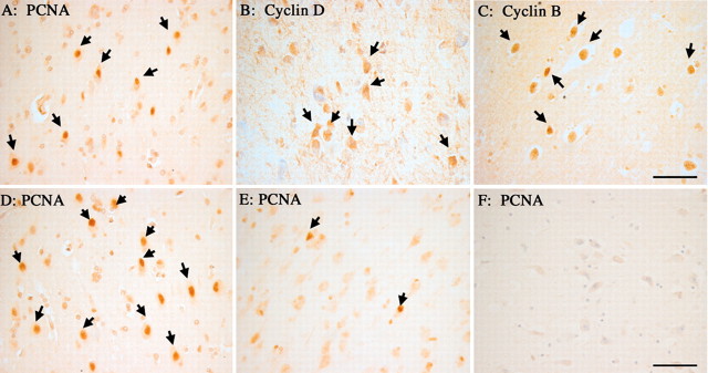
Expression of cell cycle proteins in neurons of the entorhinal cortex in MCI cases. All of the same markers found in the hippocampus are also observed in this region of temporal cortex. PCNA is found in some neuronal nuclei of Layer V of the entorhinal cortex (A). As illustrated here for neurons in layer II, neurons were also found that expressed cyclin D (B) and cyclin B1 (C). Among the different MCI cases, there was variation in the levels of cell cycle protein expression. In some cases, positive cells appeared in clusters consisting of many neurons (D), whereas in other cases only isolated immunopositive neurons were found (E). As in hippocampus, little or no PCNA staining is found in age-matched controls (F). Scale bars, 25 μm.

