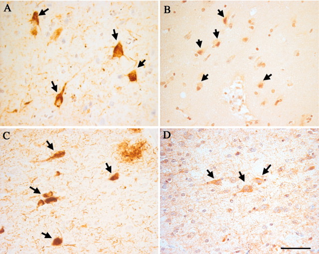
Fig. 6.
Spatial association of hyperphosphorylated tau protein and cell cycle markers in the MCI entorhinal cortex. In this series of near-neighbor sections, the monoclonal AT-8 antibody reveals the location of hyperphosphorylated tau protein in intraneuronal tangles in layer V (A) and layer II (C). In nearby sections, immunostaining for cyclin B1 (B) and cyclin D (D) shows that the cell cycle-positive neurons are closely associated with these NFT-bearing neurons. Scale bar, 25 μm.

