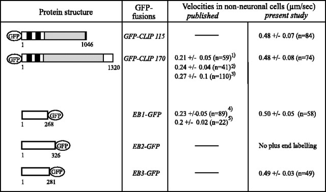
Fig. 3.
Velocities of GFP+TIP fusion proteins in non-neuronal cells. COS-1 cells, expressing the indicated GFP+TIP fusion proteins, were monitored at 37°C on a ZeissLSM510 confocal microscope and analyzed for GFP+TIP velocities. Only cells expressing low levels of the fusion proteins were investigated. For comparison, previously published values are indicated [1Perez et al. (1999); 2Akhmanova et al. (2001); 3Komarova et al. (2002a);4Mimori-Kiyosue et al. (2000); 5Morrison et al. (2002)]. MT-binding domains (black bars) and coiled-coil regions (gray bars) are indicated in the CLIPs. The microtubule-binding motif has not been identified clearly in EB1-related proteins.

