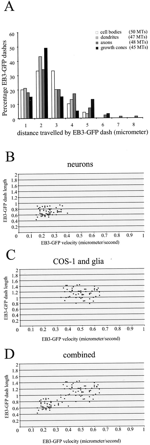
Fig. 6.

Distances traveled by EB3-GFP dashes in transfected neurons. A, Hippocampal neurons were transfected with EB3-GFP, and the cells were analyzed 3–4 d later by confocal microscopy in a 37°C chamber. The distance that individual EB3-GFP dashes could be followed was measured in different neuronal compartments (cell bodies, dendrites, axons, and growth cones). The total number of individual MTs that were counted is indicated. In all compartments the distance traveled by EB3-GFP dashes is comparable.B–D, The average speed and length of a selected number of EB3-GFP dashes derived from different measurements in neurons (B), COS-1 cells and glia (C), and the combined data (D) are plotted.
