Fig. 2.
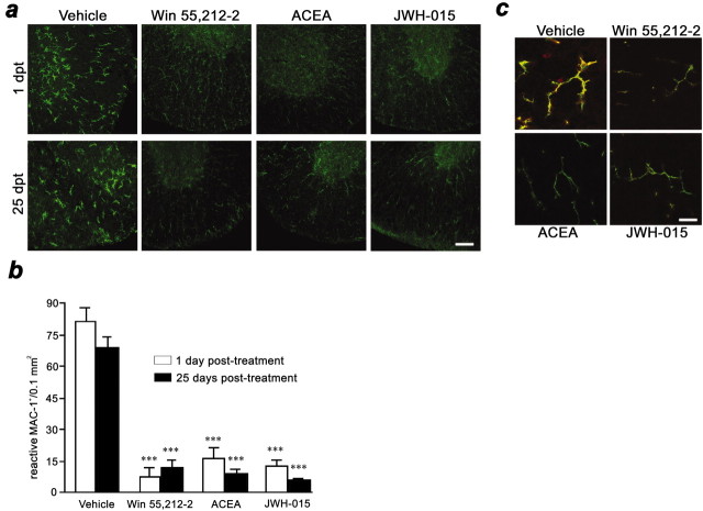
Cannabinoids inhibit microglial activation.a, Confocal images with constant laser beam and photodetector sensitivity of microglia/macrophages (CD11b+ cells) in ventral spinal cord sections. Microglial cells in vehicle-treated mice show a reactive morphology at 1 and 25 d after treatment. In contrast, WIN 55,512–2, ACEA, and JWH-015 treatments markedly inhibit reactive morphology of microglia at 1 and 25 d after treatment. Scale bar, 50 μm. b,Treatment with cannabinoid agonists reduce the number of reactive microglial cells in the spinal cord at 1 and 25 d after treatment (***p < 0.001 vs vehicle). c,Confocal images with constant laser beam and photodetector sensitivity of microglial MHC class II antigen expression. CD11b is shown ingreen, and the MHC class II complex is shown inred. Note antibody colocalization (yellow) in vehicle-treated mice and the presence of cells other than microglia expressing MHC class II molecules. Cannabinoids abrogate microglial MHC class II expression. Images are representative of 1 and 25 d after 10 d cannabinoid treatment. Scale bar, 20 μm.

