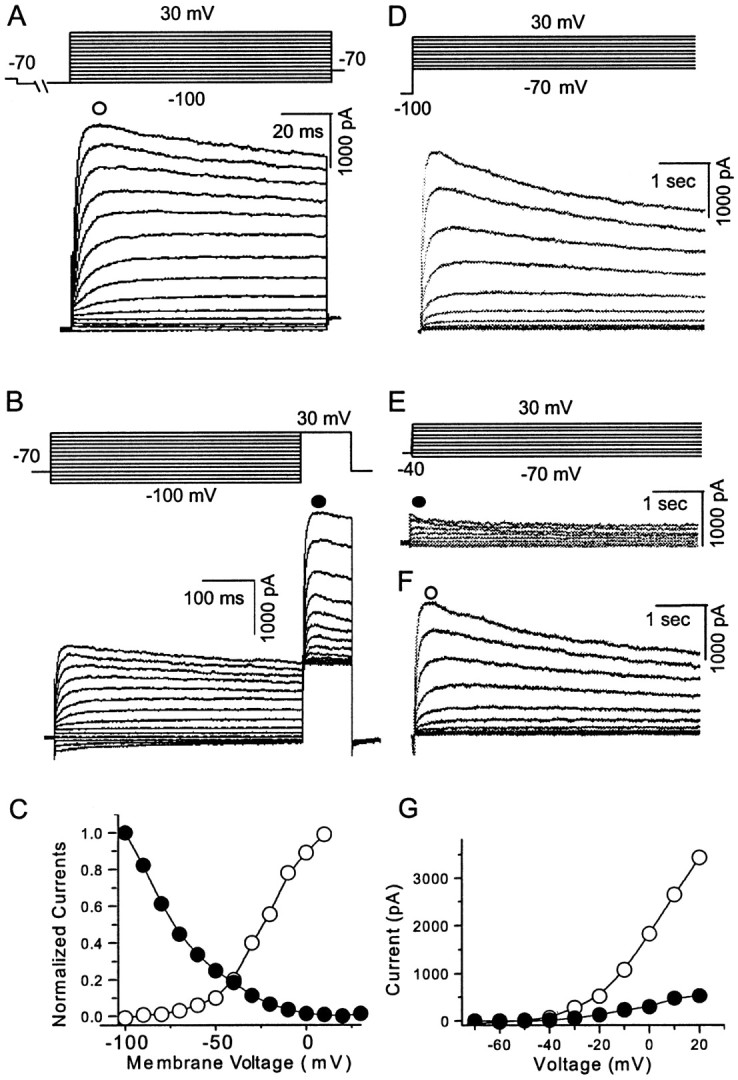
Fig. 1.

Whole-cell VGKCs in acutely dissociated mPFC pyramidal neurons. A, After holding the membrane potential at −70 mV and administering a 2 sec prepulse to −100 mV, test protocols from −100 to 30 mV with 10 mV increments elicited the whole-cell currents (n = 16 of 20).B, Whole-cell VGKCs could be partially inactivated by a depolarizing prepulse. After a holding potential at −70 mV and a 600 msec prepulse from −100 to 30 mV (10 mV increments), 100 msec test steps to 30 mV elicited currents showing a (pseudo) steady-state inactivation (n = 6). C, Current was measured at the time point indicated as an open orfilled circle in A and B. The activation and inactivation observed in A andB were plotted as I–V curves.D, Whole-cell VGKC was elicited by a similar activation protocol but with longer time course (4 sec) (n = 5). E, After the holding potential at −40 mV, the same test steps as in D elicited currents showing little inactivation. This current is operationally termedIK (n = 5).F, A slowly inactivating current was obtained by subtraction of traces in E from D. This slowly inactivating component is operationally termedID. G,I–V curves of IK andID indicate that both currents begin to activate at approximately −40 mV (n = 5). Currents were measured at the time points indicated byopen or filled circles inE and F.
