
Fig. 3.
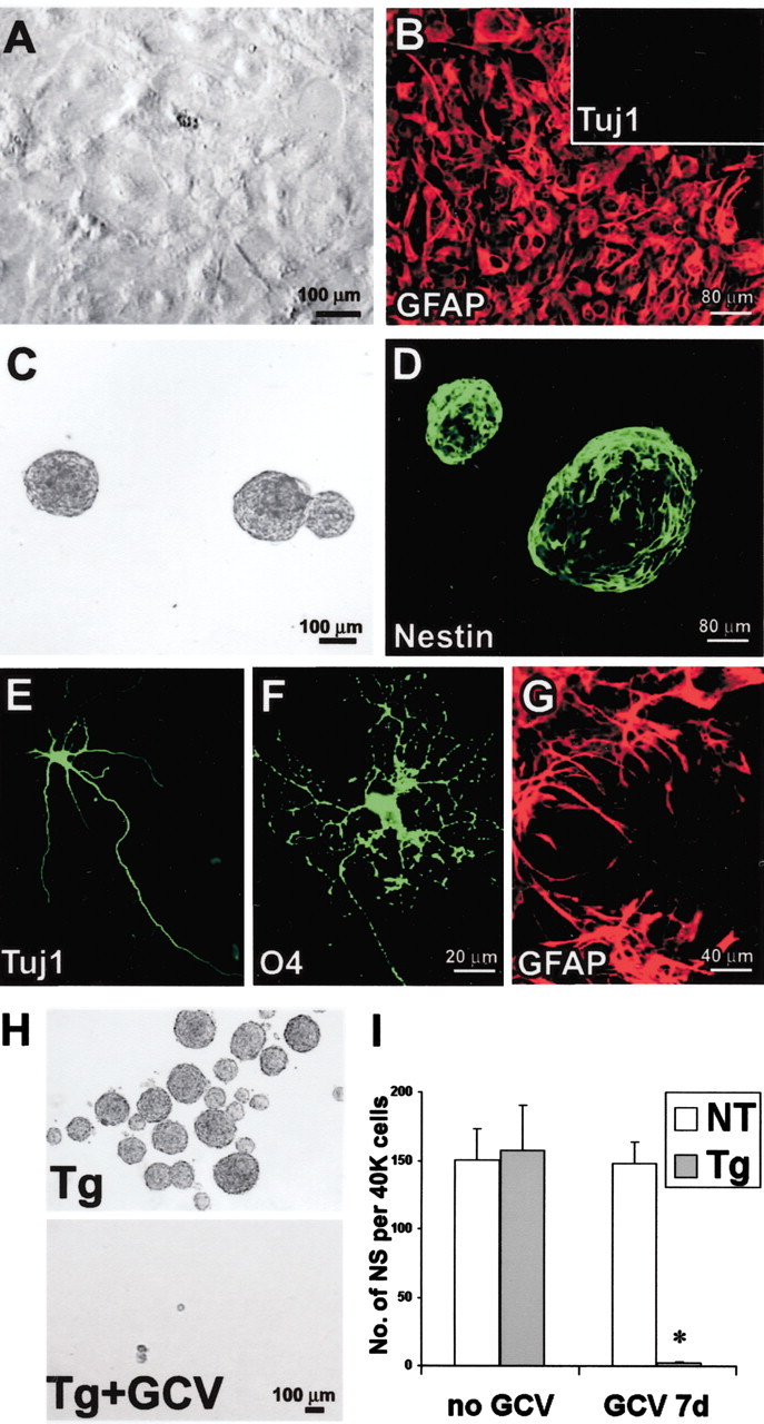
GFAP-expressing cells are required to form multipotent neurospheres from primary astrocyte cultures prepared from P1 GZs. A, Phase-contrast image of primary astrocytesin vitro showing high density of cells that have reached confluence after 21 DIV. B, Immunofluorescence of primary astrocyte culture at cell confluence after 21 DIV. Most cells stain for GFAP (red). No cells stain for Tuj1 (inset). C, Live floating neurospheres prepared from primary astrocyte culture. D, Fixed floating neurospheres prepared from primary astrocyte culture and stained for nestin (green). E–G, Immunofluorescence of chemical markers for neurons (Tuj1,E), oligodendrocytes (O4,F), and astrocytes (GFAP, G) shows cells of all three types derived by differentiation of neurospheres prepared from primary astrocyte cultures. H, Phase-contrast images of live floating neurospheres prepared from primary astrocyte cultures from GFAP-TK transgenic (Tg) mice in the absence or presence of GCV. I, Graph shows mean ± SEM number of neurospheres (NS) formed per 40,000 cells prepared after passage of primary astrocyte cultures from nontransgenic (NT) or transgenic mice in the presence or absence of GCV. GCV completely prevented the growth of spheres from transgenic mice. n = 3–5 separate cultures prepared from different mice. *Significantly different from nontransgenic or non-GCV-treated mice (p < 0.001; ANOVA plus post hoc pairwise analysis).