
Fig. 5.
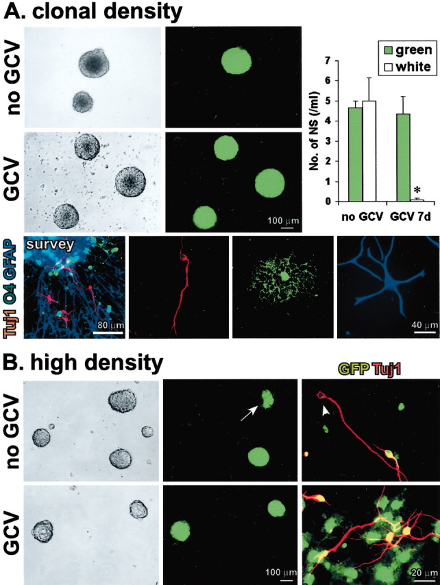
GFAP-expressing cells are not merely support cells, and their ablation is not nonspecifically toxic to neurosphere formation. A, Phase-contrast and fluorescent images of the same live floating neurospheres prepared at clonal cell density (1000 cells/ml; 500 green cells plus 500 white cells) from primary P1 astrocyte cultures with mixed single-cell suspensions of GFP and GFAP-TK cells. Without GCV, both GFP-only (green) and GFAP-TK-only (white) spheres are present. There are no mixed GFP plus GFAP-TK (green and white) spheres. In the presence of GCV, only GFP (green) spheres are present. Graph shows mean ± SEM number of green or white neurospheres (NS) formed per 1000 cells in the presence or absence of GCV. n = 3 separate cultures prepared from different mice for each value. *Significantly different from non-GCV-treated or non-TK-expressing mice (p< 0.01; ANOVA plus post hoc pairwise analysis). Tricolored immunofluorescence of markers for neurons (Tuj1,red), oligodendrocytes (O4,green), and astrocytes (GFAP, blue) shows that on differentiation, clonal spheres gave rise to different cells expressing markers of all three neural cell types, as depicted in a triple-labeled survey and details of individual cells of each type.B, Phase-contrast and fluorescent images of the same live floating neurospheres prepared at high cell density (40,000 cells/ml) from primary P1 astrocyte cultures with mixed suspensions of GFP and GFAP-TK cells. In the absence of GCV, three types of spheres formed: GFP only (green), GFAP-TK only (white), and mixed GFP and GFAP-TK (green and white;arrow). In the presence of GCV, only GFP-only (green) spheres formed. Merged double-labeled images of immunocytochemistry for Tuj1 (red) plus GFP (green) show that differentiation of spheres grown without GCV gave rise to both GFP-positive (red plus green =yellow) and GFP-negative (red,arrowhead), Tuj1-positive neurons, whereas after differentiation of spheres grown with GCV, all Tuj1-positive neurons were also GFP positive (red plusgreen = yellow).