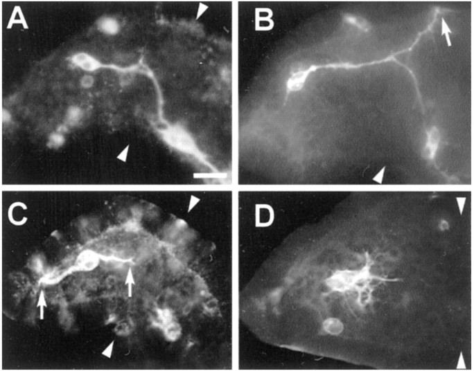
Fig. 4.
Ti1 pioneer growth cones occasionally misproject up the chemorepulsive gradients of Sema 2a. A, A representative example of the Ti1 pioneer projection into the CNS.B, A representative example of the dorsal projection error phenotype. The arrow denotes the misguided Ti1 pioneer growth cone migrating within the dorsal trochanter; the sibling Ti1 growth cone has successfully completed its projection into the CNS. C, A representative example of the distal projection error phenotype. These errors were typically direct projections of the distal Ti1 cell body axon into the extreme distal tip of the limb bud. Although it is not unusual for the distal neuron to initiate an axon at its distal pole (Lefcort and Bentley, 1989), it typically reorients its axon to grow proximally. Arrowsdenote proximal and distal projecting growth cones of the sibling Ti1 cell bodies. D, A representative example involving the failure to extend a single axon (termed the multiple short axon phenotype). Typically, the Ti1 pioneer cell bodies displayed multiple short axons projecting radially from around the cell body periphery. We interpret this phenotype as a guidance error, because normally when Ti1 neurons initiate several axons, most of them retract after extending a short distance proximally along the epithelium (O'Connor et al., 1990); however, it cannot be ruled out that this phenotype may indicate a role for Sema 2a in axonogenesis as well as pathfinding. A–D, Arrowheadsdenote the trochanter limb segment. Scale bar: A, B, 60 μm; C, D, 50 μm.

