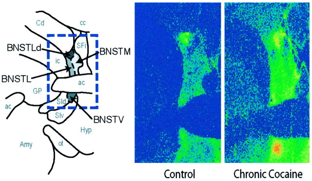
Fig. 3.
Comparison of [3H]nisoxetine binding in the BNST of representative control and chronic cocaine self-administration monkeys. Shown are coronal sections through the BNST. At left is a schematic diagram depicting the spatial organization of subregions within the BNST in which the densities of [3H]nisoxetine binding sites were quantified. At right are color-coded transformations of autoradiograms of [3H]nisoxetine binding sites in the BNST of a food control (left) and chronic (100 d) cocaine monkey (right). Each colorrepresents a range of values expressed as femtomoles per milligram of wet weight tissue. Higher levels of [3H]nisoxetine binding are visible throughout the BNST of the chronic cocaine monkey.ac, Anterior commissure; amy, amygdala;cc, central commissure; Cd, caudate nucleus; GP, globus pallidus; Hyp, hypothalamus; ic, internal capsule; ot, optic tract; SFi, septofimbrial nucleus;SId, dorsal substantia innominata; SIv, ventral substantia innominata.

