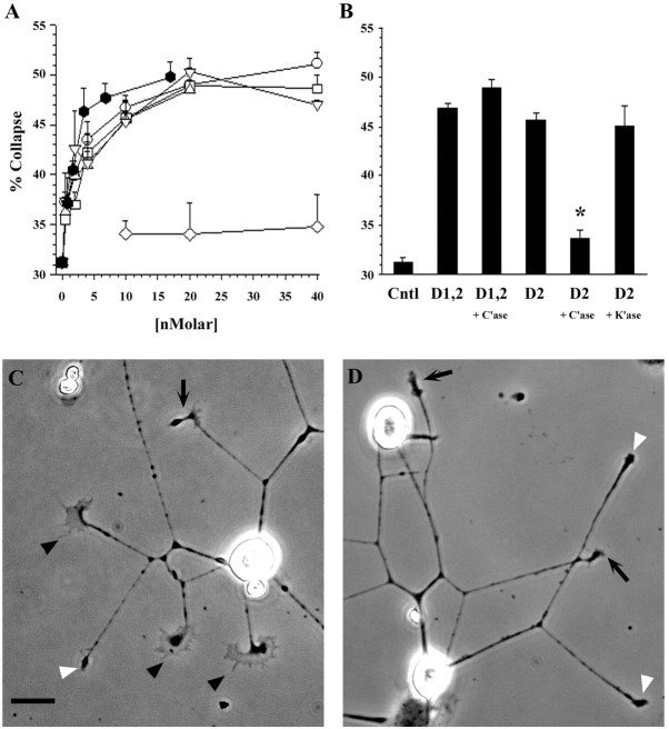
Fig. 4.
Domain-specific NG2 proteins induce the collapse of DRG neuron growth cones. DRG sensory neurons were grown on laminin for 7–8 hr and then treated with varying concentrations of NG2 fusion proteins for 30 min, fixed, and analyzed as described in Materials and Methods. A, Dose–response curves showing the collapsing activity of the different NG2 proteins. The percentage of growth cones having a collapsed morphology for each condition (y-axis) is plotted against the concentration of the test protein (x-axis): D1-Fc (■), D2-Fc (▵), D1,2-Fc (○), D3-Fc (▿), ECD-myc/his (⬡), and MUC-Fc (⋄).B, Effects of treatment of D2-containing fusion proteins with GAG lyases. D2-Fc and D1,2-Fc (10 nm) were treated with the indicated GAG lyases as described in Materials and Methods and then analyzed for growth cone-collapsing activity.C'ase, Digestion with chondroitinase ABC digestion;K'ase, digestion with keratanase. All conditions were significantly different from no-protein controls (p < 0.0001, ANOVA and Scheffé'spost hoc test), except C'ase-treated D2-Fc, which was not significantly different from controls. Values shown inA and B are mean ± SEM of two to four separate experiments. In all experiments, at least 100 growth cones were scored in each of duplicate wells per condition tested.C, D, Representative DRG neurons grown under control conditions (C) and fusion protein-treated conditions (D; D3-Fc, 20 nm). Thefilled arrowheads point to spread growth cones; theopen arrowheads point to collapsed growth cones; and thearrows point to unscored growth cones. Scale bar, 20 μm.

