
Fig. 6.
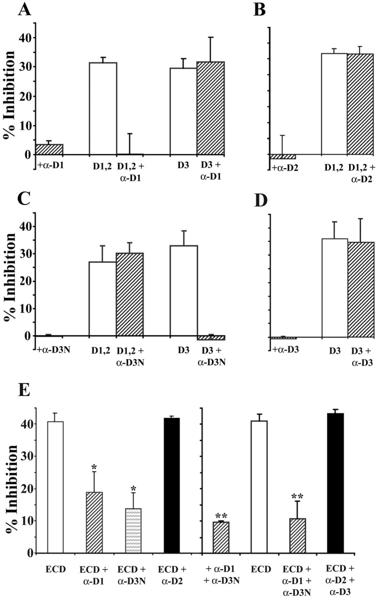
Monoclonal antibodies block neurite growth inhibition caused by the NG2 fusion proteins. Forty-eight-well tissue culture plates were coated with mixtures of L1-Fc and the NG2 fusion proteins and treated with monoclonal antibodies, and neurite lengths were measured for cerebellar granule neurons as described in Materials and Methods. L1-Fc was used at 2.5 μg/ml; D1,2-Fc (D1,2), D3-Fc (D3), and ECD-myc/his (ECD) were used at 20 nm. All the monoclonal antibodies were used at 5 μg/ml. In all panels, neurite growth on substrates coated with L1-Fc alone was measured and used as 0% inhibition and is not shown on the graphs. As shown in A and C, the anti-D1 and anti-D3N antibodies completely block neurite growth inhibition induced by their respective antigens but have no effect on the neurite growth inhibition induced by nonreactive fusion proteins. B, D, The anti-D2 and anti-D3 antibodies do not reverse the growth inhibition induced by their corresponding antigenic fusion proteins. When ECD-myc/his was used as the substrate, anti-D1 or anti-D3N treatment each only partially reversed growth inhibition (E,left). However, when both the anti-D1 and anti-D3N antibodies were used together to treat the ECD substrate, neurite lengths were comparable with those on L1-coated substrates also treated with both antibodies (E, right). Treatment of the ECD-coated substrates with anti-D2 or anti-D2 together with anti-D3 has no effect on the inhibitory activity of the ECD. Data shown are mean ± SEM from at least three independent experiments. In each experiment, neurite lengths were measured from at least 50 neurons for each condition. *Value significantly different from ECD (p < 0.001; Student's ttest) but not significantly different from each other (p > 0.1; Student's ttest); **value significantly different from ECD (p < 0.001; Student's ttest) but not significantly different from each other (p > 0.4; Student's ttest).