
Fig. 8.
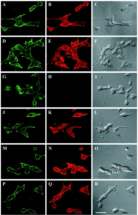
Anti-D3N antibody has limited binding to NG2 when it is expressed on the cell surface. 293-NG2 (A–L) and 293-D3 (M–R) cells were immunofluorescently stained with anti-NG2 antibodies as described in Materials and Methods. The following antibodies were used:A, D, G, J, M, P, rabbit anti-NG2; B, monoclonal anti-D1; E, monoclonal anti-D2; H, N, monoclonal anti-D3N; and K, Q, monoclonal anti-D3. Each monoclonal antibody was used at 1 μg/ml, and imaging exposure times were identical for each panel. Thethird vertical column shows the cells using DIC optics. Although anti-D1, anti-D2, and anti-D3 stain the 293-NG2 cells robustly, the signal from anti-D3N staining is significantly weaker. Anti-D3N stains the 293-D3 cells as well as does anti-D3. Scale bar, 50 μm.