Fig. 1.
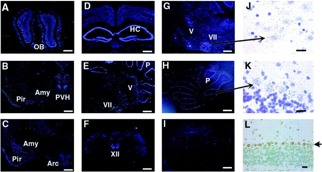
Expression of SRC-1 mRNA in the adult mouse brain.In situ hybridization was performed with35S-labeled SRC-1 antisense RNA probe. Coronal (A–D, F,L) and sagittal (E,G–K) brain sections were prepared from 10-week-old WT mice (A–H,J, K). As a negative control,in situ hybridization was performed under identical conditions on the sagittal section (I) prepared from the cerebellum region of SRC-1−/−mice of the same age. A–I andJ–L were taken under dark-field and bright-field lighting conditions, respectively. J andK were stained with hematoxylin after in situ hybridization, and their images were taken at higher magnifications from the corresponding areas indicated inG and H. Arrows indicate the facial nucleus in J and Purkinje cells inK and L. In L, SRC-1 protein (brown color) was specifically detected in the nuclei of Purkinje cells by immunostaining. Scale bars:A–I, 600 μm;J–L, 25 μm. OB, Olfactory bulb; Amy, amygdala complex;Pir, piriform cortex; PVH, paraventricular nucleus of the hypothalamus; Arc, arcuate nucleus; HC, hippocampus; P, Purkinje cell layer; V, trigeminal nucleus;VII, facial nucleus; XII, hypoglossal nucleus.

