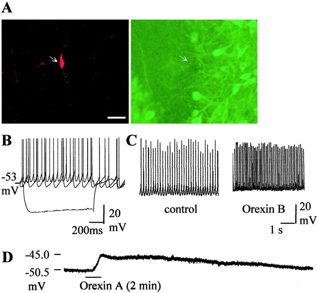
Fig. 2.
Presumed GABAergic neurons with high spontaneous firing rate are excited by orexins. A, Double stainings of biocytin-filled neuron (red) and TH-immunoreactive neurons (green). This neuron is TH negative.Arrows indicate the position of the neuron.B, Voltage responses to current pulses (−0.3, 0, +0.1 pA). C, Chart recording of membrane potential and spontaneous action potentials of the neuron before and after application of orexin B (100 nm). D, Chart recording of the membrane potential of the neuron after application of 0.5 μm TTX. Orexin A causes depolarization of the cell.

