
Fig. 1.
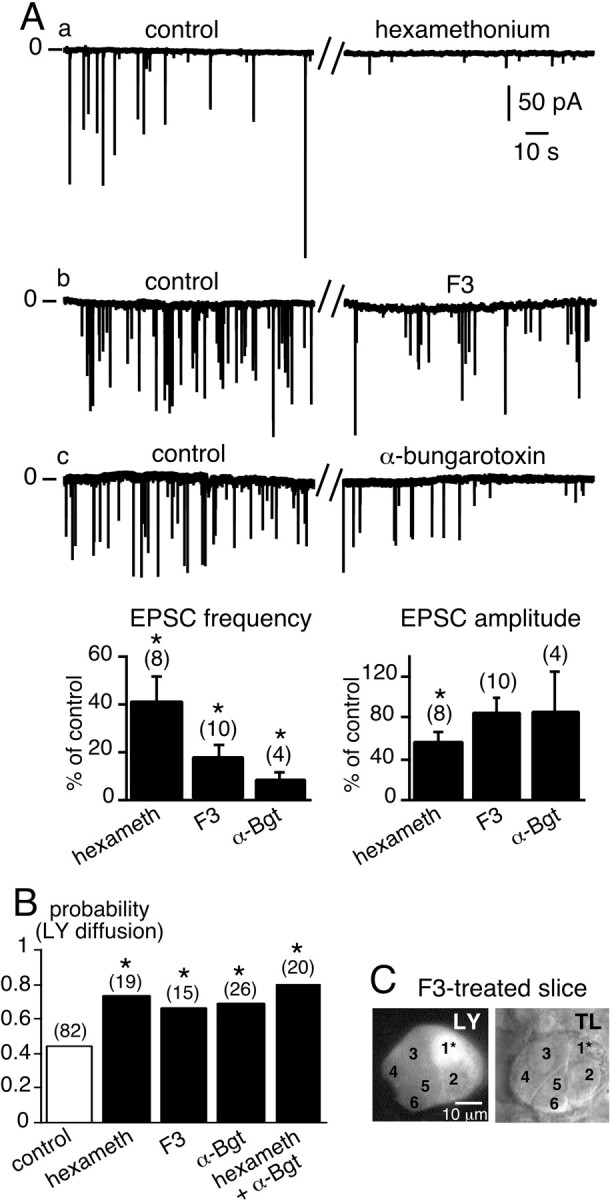
Acute impairment of synaptic activity-induced upregulation of LY diffusion between chromaffin cells. A, a–c, Spontaneous EPSCs recorded in chromaffin cells voltage-clamped at −80 mV in normal saline (2.5 mmexternal K+) (left traces) and in the presence of three bath-applied nAChR blockers, hexamethonium (200 μm), the oxystilbene derivative F3 (150 nm), and α-bungarotoxin (1.5 μm) (right traces). The histograms summarize the effects of the blockers on EPSC frequency and amplitude. *p < 0.01 compared with control values.B, Histograms illustrating the increase in the probability of LY diffusion between chromaffin cells in slices treated with nAChR blockers. The number of recorded cells for each experimental condition is indicated in parentheses. *p < 0.01 compared with control values. C, Example of widespread LY diffusion within a cell cluster in a F3-treated slice. TL, Transmitted light image. Five neighboring chromaffin cells in the same optical plane were labeled with LY after dye injection into the cell 1*.