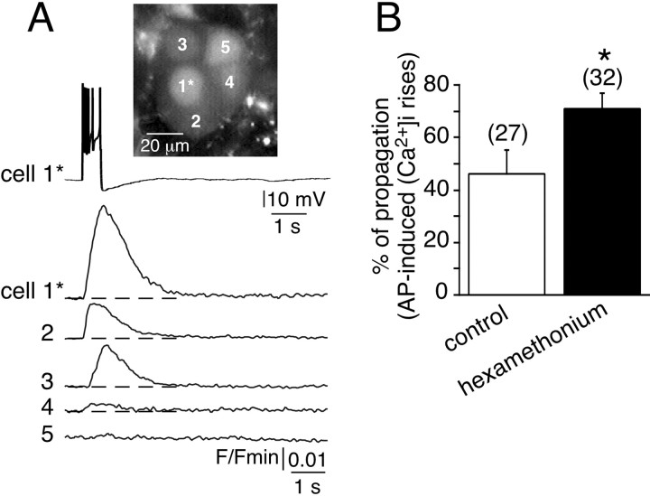
Fig. 2.
Propagation of action potential-induced [Ca2+]i rises between chromaffin cells in hexamethonium-containing saline. A, Electrical activity-driven multicellular [Ca2+]iincreases were visualized by real-time scanning laser confocal imaging (120 images per second, averaging 4 frames) in five chromaffin cells loaded with Oregon Green 488 BAPTA-1 as the Ca2+-sensitive fluorescent probe. The adrenal slice was continuously perfused with 200 μm hexamethonium (for at least 30 min before recording). The plots of relative Oregon Green 488 BAPTA-1 emission changes show a [Ca2+]i rise in either the stimulated cell (1*, burst of action potentials triggered by an injection of a 500 msec depolarizing current) or three nearby cells (cells 2–4). Note that cell 5 remained silent. Dotted lines indicate the baseline.B, Histogram illustrating the percentage of cell fields in which the [Ca2+]i rise was propagated to adjacent cells in control and hexamethonium-treated slices. *p < 0.01 compared with control.

