
Fig. 5.
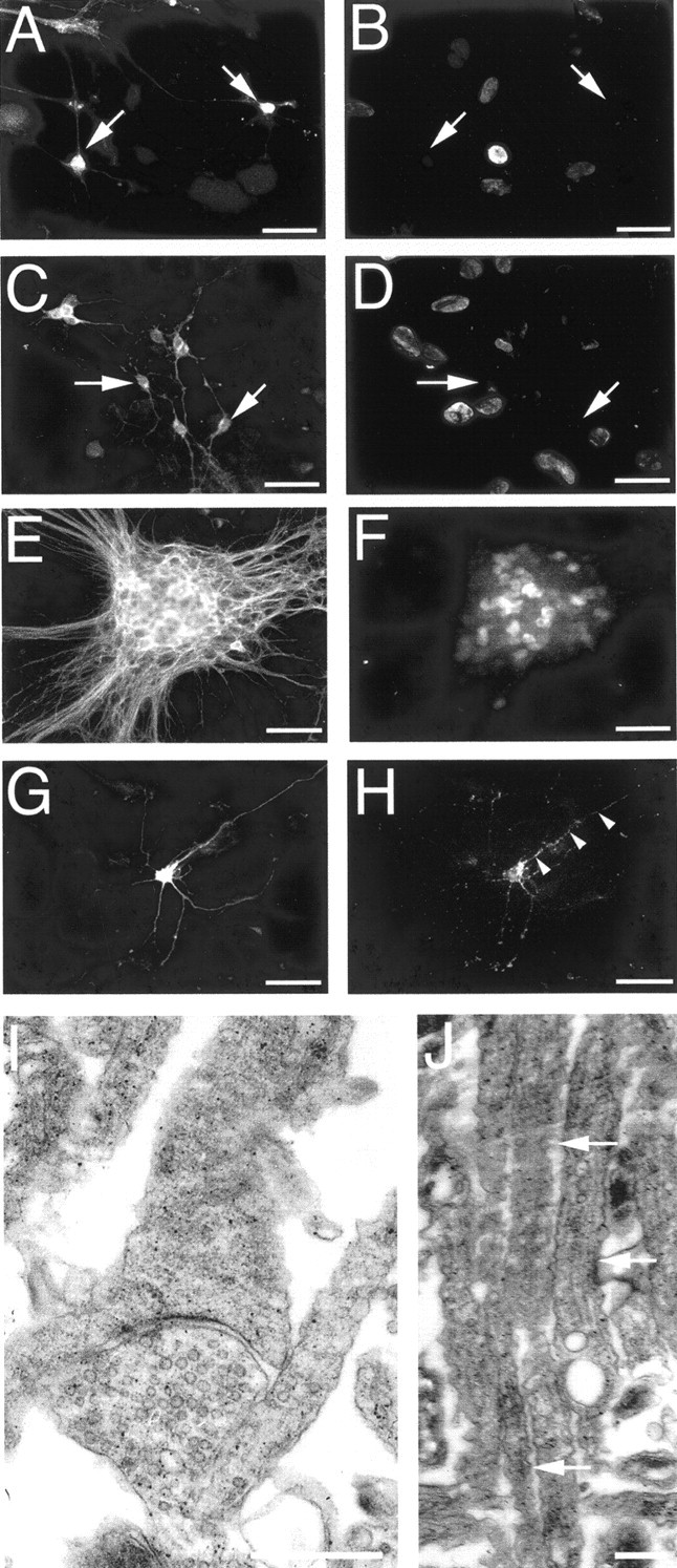
Confirmation of cortical neuron maturity. BrdU was administered to cultures at 7 (A, B) or 17 (C, D) DIV, for cell proliferation assays, and cultures were grown to 21 DIV. Neuron-specific enolase (NSE) immunopositive neurons (arrows in A andC) lacked BrdU-immunoreactive nuclei (B,D) in double-labeling studies, indicating that they were not proliferating. Double labeling for tau (E) and postmitotic neuronal nuclei (F) at 21 DIV demonstrated that aggregated neurons were not dividing. NSE-labeled neuron (G) in apposition with a profile of synaptophysin-immunoreactive puncta (arrows depict examples inH), demonstrating that neurons had abundant synaptic connections. The presence of synapses was confirmed with electron microscopy (I). J, Electron microscopy demonstrating that axons within axonal bundles were not myelinated (arrows depict examples). Scale bars:A–H, 30 μm; I, 2 μm;J, 1 μm.