
Fig. 7.
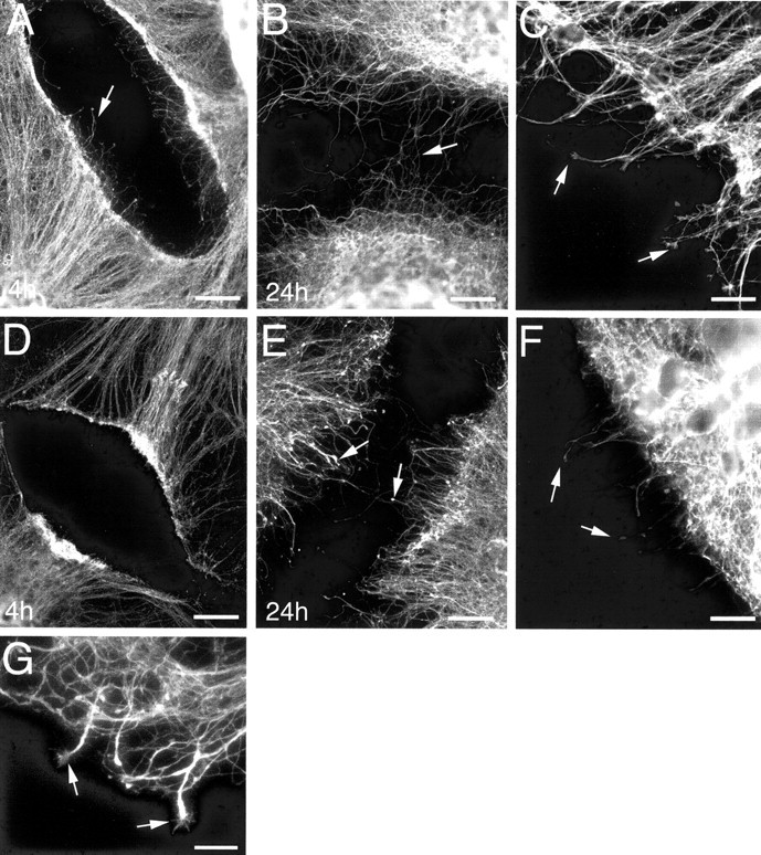
Immunofluorescence labeling for tau, demonstrating the effect of taxol exposure on postinjury axonal sprouting. Times shown on each image indicate hours of vehicle or taxol exposure after injury. Vehicle-treated cultures demonstrated an extensive and progressive sprouting response after axonal transection, with several sprouts crossing the lesion site by 24 hr after injury (A, B). Postinjury sprouts elaborated from vehicle-treated cultures were typically slender with expanded growth cone-like distal domains (arrows in C). Limited postinjury axonal sprouting was demonstrated in taxol-treated cultures up to 24 hr after injury (D, E), and bulb-like structures formed at the tips of sprouts (arrows inF). Arrows in A, B,D, and E depict examples of postinjury sprouts. C and F were captured at 4 and 14 hr after injury, respectively. Taxol washout, after 4 hr of postinjury taxol exposure, resulted in increased sprout growth across injury sites, and some sprouts gained growth cone-like structures at their tips (arrows in G). Scale bars: A,B, D, E, 60 μm;C, F, 20 μm; G, 10 μm.