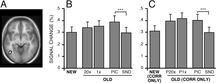
Fig. 5.
Left inferior temporal cortex near BA 19/37 increased activity during retrieval of picture content. Format is similar to that of Figure 3. A, Left BA 19/37 region near the anterior occipital sulcus (z = −8).B, Magnitude estimates of signal change in BA 40/39 for each condition. C, Magnitude estimates when categorized by response accuracy.

