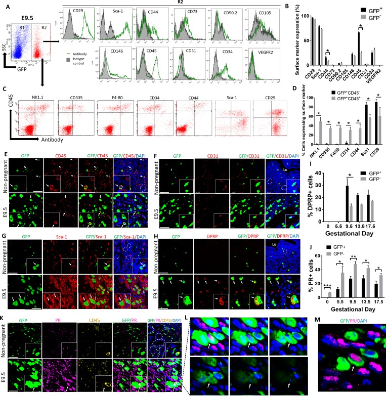
Fig 2. Characterization of uterine BMDCs throughout pregnancy.
(A) Flow cytometry analysis of BMDCs in uterine implantation site on E9.5. Cells gated in R2 are BM derived (GFP+) uterine cells, while cells gated in R1 are non–BM-derived resident (GFP−) uterine cells. Histograms represent GFP+ cells (R2) from E9.5 implantation sites that are stained with the indicated antibodies (green line) and respective isotype controls (filled) (n = 4). (B) Quantification of percentage of BM-derived (GFP+) and non–BM-derived (GFP−) uterine cells expressing the various cell surface markers shown in (A) (n = 4), *p < 0.05. (C) Flow cytometry analysis of E9.5 BM-derived (GFP+) uterine cells (R2) using CD45 in combination with various surface marker antibodies. (D) Quantification of surface markers shown in (C), on BM-derived nonhematopoietic decidual cells (GFP+CD45−) and BM-derived hematopoietic cells (GFP+CD45+) (n = 4–5), *p < 0.01. (E, F, G, H, K) Immunofluorescence photomicrographs of E9.5 mesometrial decidua sections or nonpregnant mice uteri sections demonstrating co-staining of GFP+ BMDCs (green) with (E) CD45 (red), (F) CD31 (red), (G) Sca-1 (red), (H) DPRP (red), (K) progesterone receptor (PR) (pink), and CD45 (yellow). Sections were counterstained with DAPI showing nuclei (blue). Insets show higher magnification photomicrographs. White arrows point to BMDCs colocalizing with their respective markers. White dashes are encircling endometrial glands. Scale bars, 50 μm. (L and M) A z-stack series (L) and a 3D image (M) of the inset from (K) demonstrating a single BM-derived GFP+ cell co-expressing PR but negative for CD45 (white arrow). (I) Quantification of BMDCs (GFP+) or non-BMDCs (GFP−) positive for DPRP throughout gestation (n = 3). *p ≤ 0.01. (J) Quantification of BMDCs (GFP+) or non-BMDCs (GFP−) positive for PR throughout gestation (n = 3–5). *p ≤ 0.05, **p ≤ 0.01, ***p ≤ 0.001. In all panels, bar graphs represent mean ± SEM. See also S3–S5 Figs. Underlying data are available in S1 Data. BM, bone marrow; BMDC, BM-derived cell; DPRP, decidual prolactin-related protein; GFP, green fluorescent protein; PR, progesterone receptor; Sca-1, stem cell antigen-1; SSC, side scatter; VEGFR2, vascular endothelial growth factor receptor 2.

