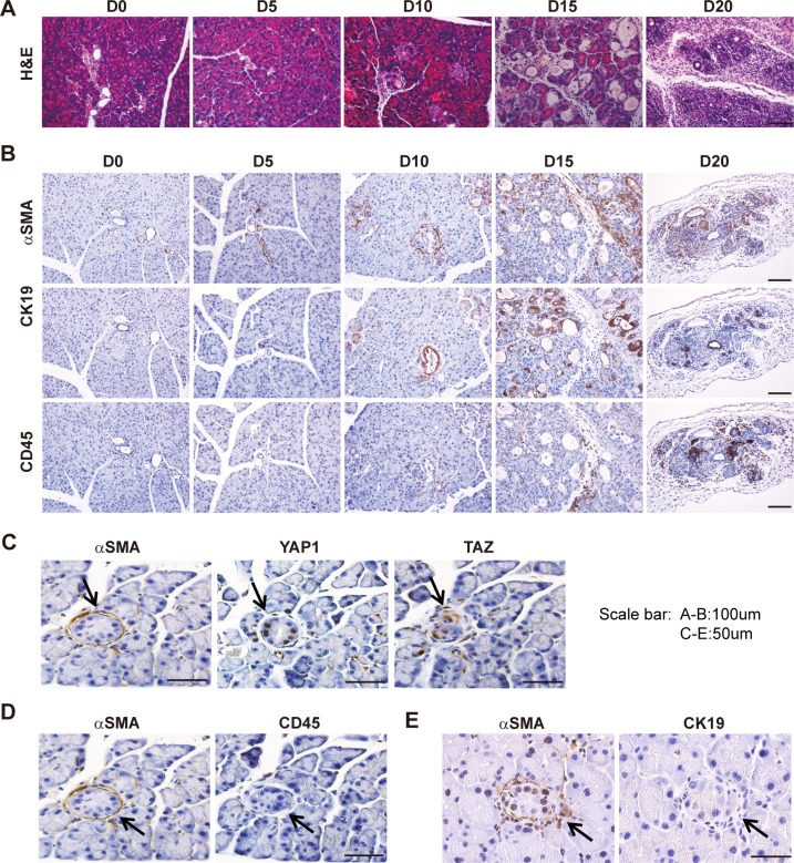
Fig 4. Lats1/2 deletion in acinar cells results in PSC activation.
PL mice were injected once with 180 mg/kg of TAM; pancreata were collected and embedded at Day 0, Day 5, Day 10, Day 15, and Day 20 after injection, respectively (n = 4–6). (A) Morphological changes over time (Day 0, Day 5, Day 10, Day 15, Day 20) were examined by HE staining. (B) Anti- αSMA, anti-CD45, and anti-CK19 antibodies were used to stain activated PSCs, immune cells, and ductal-like cells, respectively. (C) αSMA, YAP1, and TAZ IHC staining in consecutive sections at Day 10. (D) αSMA and CD45 IHC staining in consecutive sections at Day 10. (E) αSMA and CK19 IHC staining in consecutive sections at Day 10. αSMA, α-smooth muscle actin; CD45, cluster of differentiation antigen 45; CK19, cytokeratin 19; HE, hematoxylin–eosin; IHC, immunohistochemistry; LATS1, large tumor suppressor 1; PL, double knockout; PSC, pancreatic stellate cell; TAM, tamoxifen; TAZ, transcriptional coactivator with PDZ binding motif; YAP1, yes-associated protein 1.

