Fig. 3.

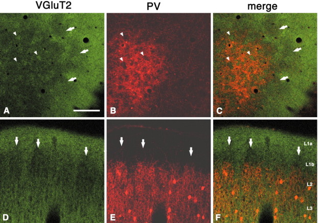
Immunohistochemistry for VGluT2 shows dense uniform staining in layer 1a but a discontinuous periodic pattern in layers 1b and 2 (A–C, tangential section;D–F, coronal section). Double labeling for VGluT2 and PV shows that VGluT2-ir dense regions in layer 2 are situated within the PV hollows. Arrowheads point to corresponding spaces (hollows for PV and dense regions for VGluT2). The complementary relationship extends into layer 1b, where VGluT2 sparse areas (arrows) can be seen above PV walls, inD–F. Scale bar (shown in A):A–C, 200 μm; D–F, 100 μm.
