
Fig. 6.
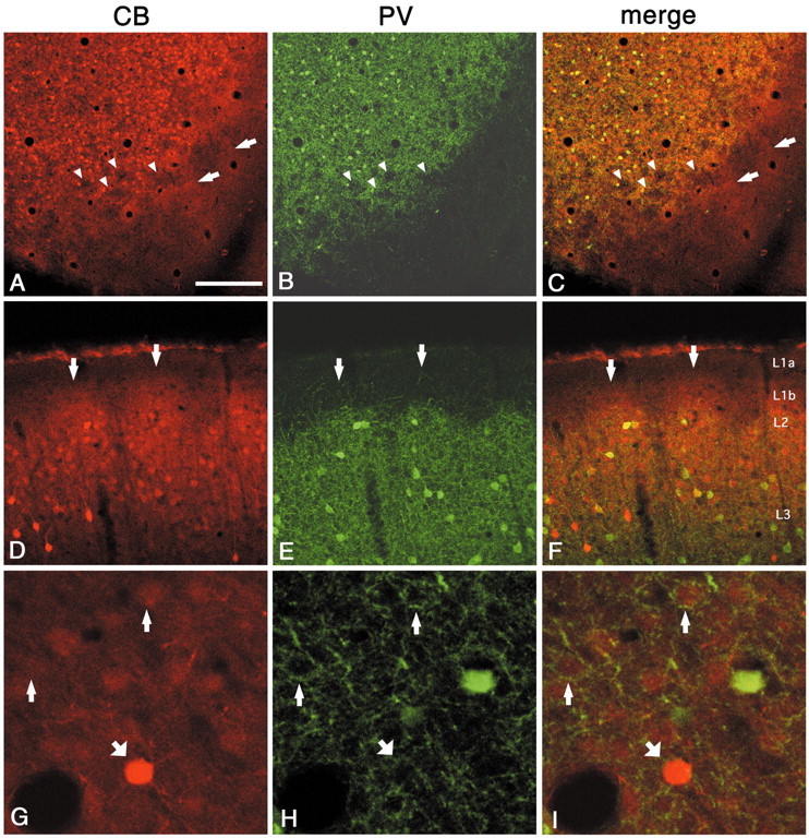
Double labeling for CB and PV demonstrates that regions dense for CB colocalize with PV in the walls (A–C, tangential section; D–F, coronal section). As with zinc, the CB pattern extends higher, into layer 1b (arrows). Arrowheads point to corresponding hollows in both markers. G–I, Higher magnification from walls (tangential section). Thin andthick arrows point, respectively, to weakly and strongly CB-ir cell bodies surrounded by basket-like PV-ir terminal puncta. Scale bar (shown in A): A–C, 200 μm;D–F, 100 μm; G–I, 30 μm.