
Fig. 7.
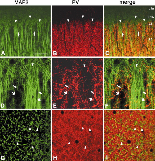
Double labeling for MAP2 and PV shows that large bundles of apical dendrites (probably from neurons from layer 3 and layer 5) lie predominantly within PV hollows (A–C,coronal section; D–F, higher magnification;G–I, tangential section). The separation between walls and hollows is less clear than in PV, and some MAP2-ir dendrites, weakly stained and forming small bundles, can be seen in the honeycomb walls (A, C, arrows). In contrast with VGluT2, MAP2 immunohistochemistry does not show any distinct pattern in layer 1b, which is uniformly filled with fine MAP2-ir particles, probably representing apical dendritic tufts. Apical dendrites from layer 2 pyramidal neurons (D–F, thinner arrows) are less frequently found to be MAP2-ir. These can be traced, however, to weakly MAP2-ir somata or unstained somata, visualized by PV-ir basket-like terminals (D–F, thicker arrows).Arrowheads point to corresponding PV hollows and MAP2-ir large dendritic bundles. Scale bar (shown in A):A–C, 100 μm; D–F, 25 μm;G–I, 100 μm.